One Model, Heart and Vessels: How MeshGrow Builds Simulation-Ready Cardiovascular Anatomy
A plain-language explainer of MeshGrow, an automated framework for constructing combined cardiac and vascular models from medical images
The Problem
To simulate blood flow through a patient’s cardiovascular system—to plan a procedure, test a device, or study how disease is progressing—you first need an accurate 3D model of that person’s anatomy. And ideally, you want the whole picture: the heart chambers pumping the blood and the arteries carrying it away.
The trouble is that building these models from a CT or MR scan is one of the slowest, most manual steps in the entire pipeline. Existing automated methods have mostly picked a side: some are good at reconstructing the cardiac chambers, others at tracing the vasculature. Very few produce a single, connected, simulation-ready model of both.
MeshGrow is our attempt to close that gap.
Why the Heart and the Vessels Fight Back
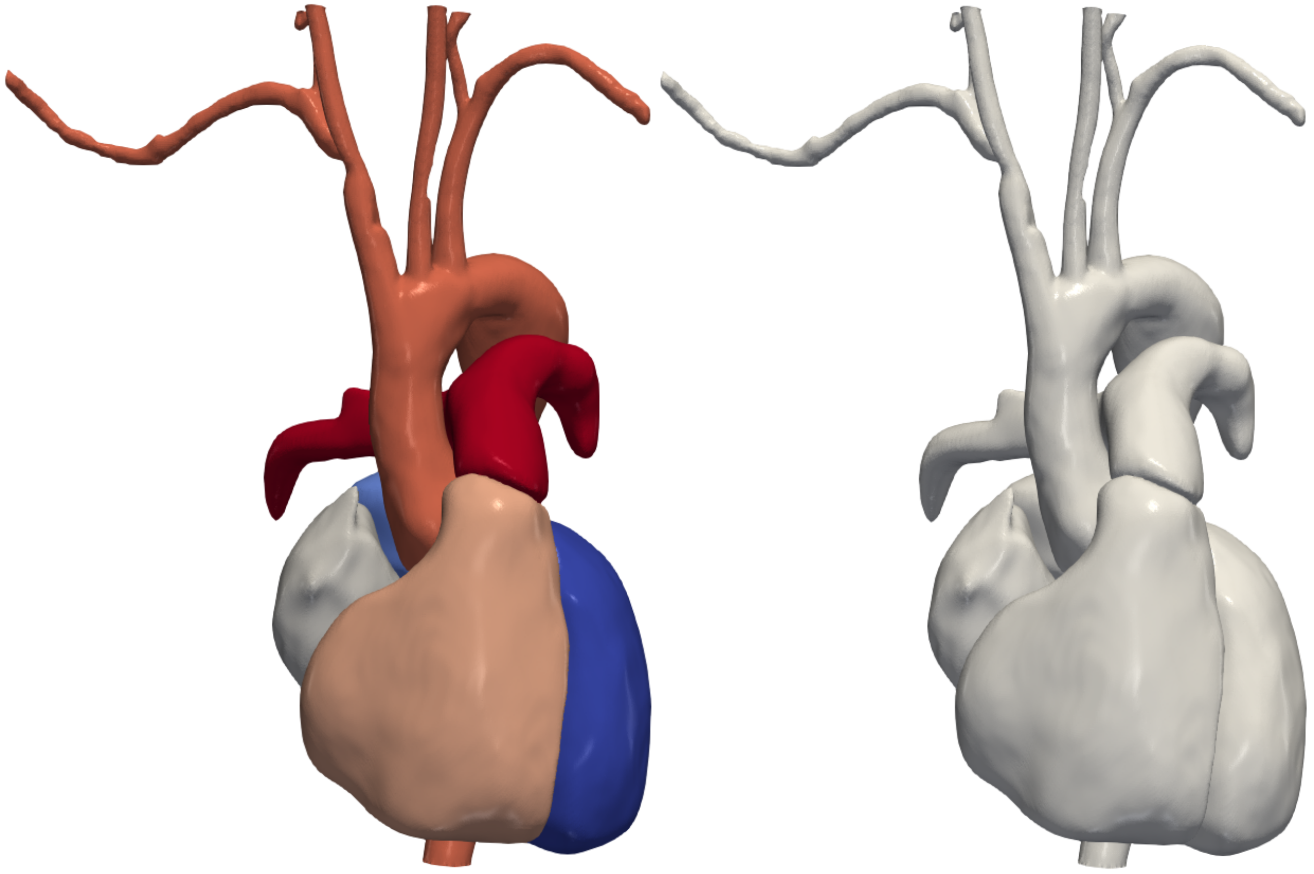
Cardiac chambers and blood vessels look nothing alike, and that is exactly the problem.
- The heart chambers are big, blob-like volumes. Their overall shape is fairly consistent from patient to patient, so a method that “knows” what a heart looks like can deform a template to fit a new scan.
- The vessels are thin, branching tubes that vary enormously between people and make up only a tiny fraction of the pixels in a scan. A rigid template can’t anticipate where every branch goes; you have to follow them.
Trying to force one technique to handle both anatomies tends to do neither well. So instead of looking for a single hammer, MeshGrow uses the right tool for each job.
The Core Idea: Two Tools for Two Anatomies
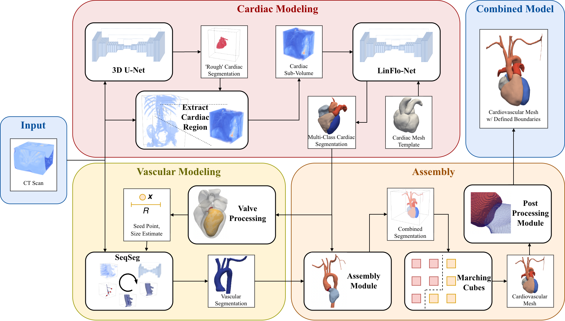
MeshGrow is a two-stage framework:
-
Mesh the heart. For the cardiac chambers, we use a template-based deep learning approach. The method starts from a mesh that already encodes what a healthy heart’s chambers look like, and deforms it to match the patient in the scan. Because the template carries prior knowledge of cardiac anatomy, this stays robust even where image quality is poor.
-
Grow the vessels out from it. For the aorta and its main sub-branches, we use a step-wise, growth-based tracer (the same family of ideas behind SeqSeg): starting near the heart, it follows each vessel locally, one small piece at a time, and assembles the branches into a connected tree.
The name says it all—we mesh the heart first, then grow the vasculature outward from it.
Stitching the Heart to the Vessels
Building two halves is only useful if they connect properly. The interesting engineering in MeshGrow is at the seam: the framework joins the cardiac mesh and the vascular mesh into a single watertight surface, and—critically—defines an aortic valve surface where the left ventricle meets the aorta.
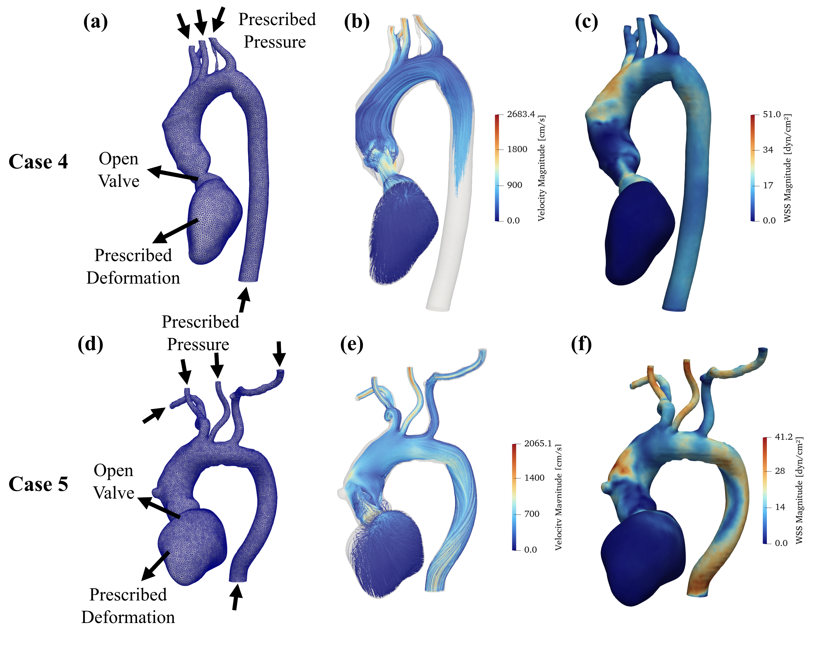
That valve surface, along with the vessel inlets and outlets, is exactly what a solver needs to apply boundary conditions: where blood enters, where it leaves, and how the valve gates flow. Without them, you have a pretty picture; with them, you have a computational domain you can actually run physics on.
What We Found
We tested MeshGrow on five CT datasets, comparing its output against state-of-the-art benchmark methods and against ground-truth models built by hand.
- Higher accuracy. MeshGrow achieved higher metric scores than the benchmark methods across the test data.
- It actually simulates. To prove the models are simulation-ready and not just visually plausible, we ran full three-dimensional computational fluid dynamics (CFD) simulations on two of the test cases—end to end, from image to hemodynamics.
- One connected model. By meshing the chambers and growing the vessels with anatomy-specific tools and then fusing them, MeshGrow returns a unified mesh with the valve and boundary surfaces already in place.
Why This Matters
Most automated modeling tools force a choice between the heart and the vessels. MeshGrow shows that you don’t have to choose: by matching the method to the anatomy—template deformation for the chambers, growth-based tracing for the vessels—you can automate the construction of a combined cardiovascular model.
That is a step toward making patient-specific simulation practical at scale: large-cohort studies and, eventually, time-sensitive clinical settings where waiting hours to hand-build a model isn’t an option.
Read the Paper
MeshGrow is published, open access, in JRSM Cardiovascular Disease:
- Paper: MeshGrow: Integrated framework for simulation-ready cardiac and vascular mesh construction from medical imaging
- Earlier conference version: Integrated Framework for Unified Cardiac and Vascular Mesh Construction from Medical Images (FIMH 2025)
- Related work: the vessel-growing idea builds on SeqSeg
This work was done with Arjun Narayanan, Fanwei Kong, and Prof. Shawn Shadden. Curious about automated cardiovascular modeling, or have thoughts on where the remaining bottlenecks are? I’d love to hear from you—reach out or leave a comment below.
Enjoy Reading This Article?
Here are some more articles you might like to read next: